About Retinal Vein Occlusion

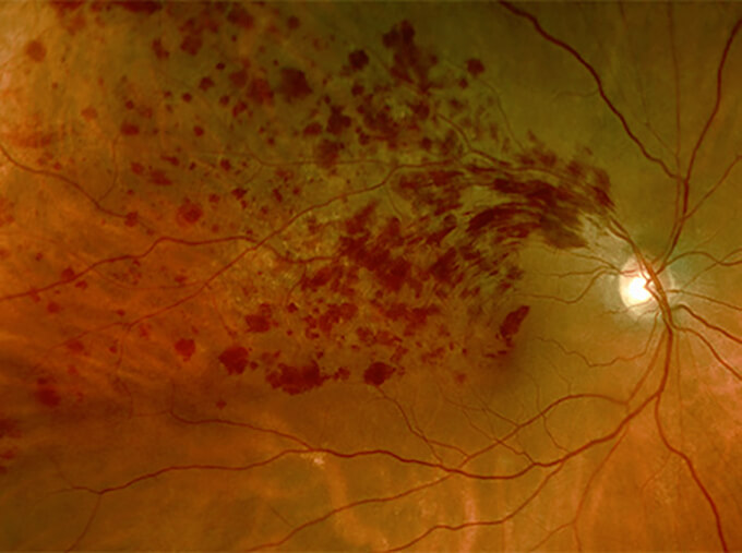
Vein occlusions are caused by a blockage of the blood flow in a vein. They are more likely to occur in older patients, and patients with a history of glaucoma, high blood pressure, and cardiovascular disease. Sometimes, vein occlusions can be associated with other systemic diseases such as blood clotting disorders. Your physician will determine if additional testing or evaluation is needed to determine the cause of the vein occlusion.
The symptoms of vein occlusion can be mild to severe depending upon the location and type of vein occlusion. Painless vision loss can occur which involves part or all of the vision. The loss of vision may be due to swelling of the retina or loss of blood flow to parts of the retina. Vein occlusions can also cause other complications such as the growth of abnormal blood vessels in the eye and high eye pressure. For these reasons, it is important to see a retinal specialist quickly if you have symptoms or are diagnosed with a vein occlusion.
Treatment is usually directed at the complications of vein occlusion, such as the swelling of the retina (“macular edema”). Treatment may involve injections of medications into the eye, such as anti-vascular-endothelial-growth-factor agents or steroids, or laser therapy. The goal of treatment is improvement of vision. Multiple treatments may be required to improve or maintain vision after a vein occlusion.
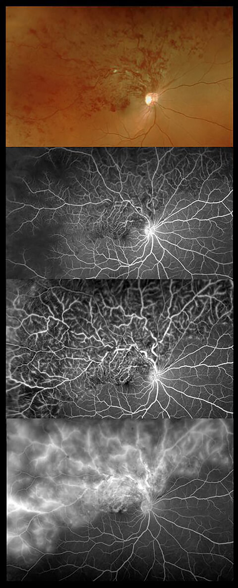
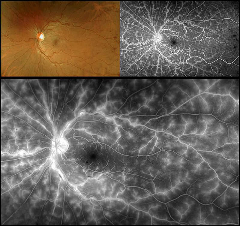
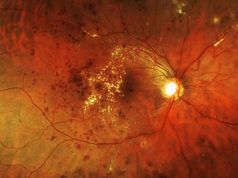
Diagnosis of vein occlusions is typically made through a combination of the dilated examination, ophthalmic photos, fluorescein angiography (where dye is injected into a vein in your arm to assess blood flow within your eye), and optical coherence tomography.
For additional information, please visit the ASRS page on branch or central retinal vein occlusions.


